Diagram of The Human Nose


Diagram of The Human Nose

A diagram illustrating the human nose generally encompasses both its external and internal components. On the outside, it displays the nasal bridge, tip, nares (nostrils), alae (the sides of the nostrils), and columella (the tissue that divides the nostrils). Internally, the nasal cavity is partitioned by the septum and contains turbinates (conchae) and sinuses.
External Nose:
Nasal Bridge: The bony upper section of the nose.
Nasal Tip: The soft, rounded extremity of the nose.
Nares (Nostrils): The two apertures that permit air to enter the nasal passage.
Alae: The curved edges of the nostrils.
Columella: The tissue that divides the nostrils.
Internal Nose:
Nasal Cavity: The area within the nose, separated into two by the nasal septum.
Nasal Septum: A structure composed of bone and cartilage that divides the left and right portions of the nasal cavity.
Turbinates (Conchae): Bony projections lined with mucous membrane that assist in warming and humidifying the air.
Sinuses: Air-filled spaces within the skull that are connected to the nasal cavity.


Diagram of the Human Nose....anatomy-and-human-blockhead.


DIAGRAM OF THE HUMAN NOSE


DIAGRAM OF THE HUMAN NOSE...


The upper air passages pertain to the respiratory organs located in the human head. These include the nose and throat (pharynx).
When breathing in, air first enters through the nose, where it is initially filtered by a hair-like structure.
A bifurcated network of blood vessels within the nasal mucous membrane serves to warm the incoming air. Additionally, mucous droplets from the nasal mucous membrane help to moisten the air. This process prepares the air for the conditions found within the lungs (pulmo). The paranasal sinuses (sinus paranasales), which are connected to the nasal cavity, also play a role in this adaptation.
From the nose, the air travels to the throat, which comprises various sections, with the esophagus and windpipe intersecting in the medial throat (mesopharynx). The inhaled air continues through the throat to the larynx (larynx) and then to the lower air passages.
In contrast to the sense of taste, the sense of smell is capable of distinguishing a far greater variety of sensations. The qualitative categorization seen in taste (bitter, sweet, sour, salty) does not apply as straightforwardly to smell. Instead, scent classes are utilized to convey the experience (pungent, sweaty, rotten, etc.).
The sensory receptors of the olfactory organ (organum olfactus) are situated at the beginning of the respiratory tracts and are approximately the size of a dime, located on the upper nasal wall and the septum.
Due to pigmentation, the olfactory region appears yellow, which contrasts with the red membrane. It remains uncertain whether this pigmentation plays a role in the sense of smell. However, it is noted that animals lacking pigmentation (albinos) do not possess a sense of smell.
The tissue within the olfactory region, containing over 10 million receptor cells, is approximately 50 µm thicker than the epithelium of the respiratory tracts. The olfactory cells feature very fine hairs (cilia), with up to 12 per cell, and are coated in mucus. Nerve processes from the olfactory cells bundle together as fibers leading to the anterior section of the olfactory cortex at the base of the frontal brain. The olfactory cells are separated from the olfactory cortex by a very thin bone known as the sieve.. The olfactory cells are separated from the olfactory cortex by very thin bone, the sieve, through which the nerve fibers pass through small openings. After appropriate processing of the olfactory information, the smell becomes known.

Olfactory cells are chemical receptors, i.e. the stimulus of the olfactory cells is the result of a chemical process on the surface of the hairs. How the molecular reaction occurs, with which hundreds of different smells are distinguished, is unclear. Gaseous substances are easier to smell. A water-soluble property increases the ability to smell because the smell molecules enter the air through evaporation.


Diagram of the Human Nose....

diagram-of-human-nose


Structure of the Nose


Structure of the Nose

The external nose is visible and is pyramidal in shape, with the root located in the upper region and the base located in the lower region. The variance in shape depends on the shape of the ethmoid bone which is an anterior cranial bone located between the eyes. The internal nose is divided into the left and right nasal cavities by the nasal septum. The internal nose also consists of the skin-lined region known as the vestibule and the mucosa-lined region known as the nasal cavity proper.

The nasal framework

The nasal or piriform aperture is situated on the bony skull and represents a bony outline of the nasal cavity, which is the beginning of the respiratory tract. The nasal pyramid is located here with each side consisting of the frontal process of the maxilla and medial to the nasal bone which is linked to the frontal bone.

The nasal cartilage

The front of the nose consists of connective tissue with embedded small pieces of cartilage. This includes the nasal septum and the nasal tip cartilage (lesser alar cartilage and lateral greater alar cartilage). Whatever impression the nose makes as an aesthetically significant element of the face – high, broad, or even crooked – is largely determined by these small cartilages and the cartilaginous portions of the nasal septum. Frequently, the bony portion of the nose is affected by midfacial fractures (e.g., a nasal bone fracture).

A common cause of nasal fractures is blunt trauma. Manual alignment through the application of strong thumb pressure with anesthesia followed by repair may be necessary to prevent lasting misalignments. Injuries that lead to functional impairments may be corrected through nasal surgery or rhinoplasty.

Internal nose

The inner part of the nose is formed by the main nasal cavity which includes the nasal concha, nasal passages (nasal meatus), and paranasal sinus. The nasal septum comprises bone and cartilage in the nose and separates the nasal cavity into two fossae, which are continuations of the nostrils and jointly form their medial border. The nasal septum consists of four structures:

Clinically, deviations of the nasal septum occur quite frequently. These deviations may be present at birth or may result from an accident. A deviated nasal septum can lead to nasal congestion and breathing difficulties which may require surgical correction. Structure of the Nose .

Structure of the Nose Video :


UNDERSTANDING NASAL ANATOMY


 UNDERSTANDING NASAL ANATOMY

A lot happens under the surface of the nose. The bone and cartilage under the skin give the nose most of its size and shape. Other structures inside and behind the nose help you breathe. Learning the anatomy of the nose can help you better understand how the nose works.

Bone. 

This supports the bridge of the nose. The upper cartilage supports the side of the nose. The lower cartilage adds support, width, and height. It helps shape the nostrils and the tip of the nose.

Skin. 

This also helps shape the nose.

Nasal cavity. 

This is a hollow space behind the nose that air flows through.

Septum. 

This is a thin wall made of cartilage and bone. It divides the inside of the nose into 2 parts.

Mucous membrane. 

This is thin tissue that lines the nose, sinuses, and throat. It warms and moistens the air you breathe in. It also makes the sticky mucus that helps clean the air of dust and other small particles.

Turbinates. 

These are curved, bony ridges on each side of the nose. They are lined with mucous membrane. They warm and moisten the air you breathe in.

Sinuses. 

These are hollow, air-filled chambers in the bone around your nose. Mucus from the sinuses drains into the nasal cavity.

 UNDERSTANDING NASAL ANATOMY VIDEO :


Physiology of the Human Nose


Physiology of the Human Nose

THE HUMAN NOSE

The human nose has several physiologic functions. As the air is inspired through the nose it is humidified and warmed by passing over the moist and warm nasal mucosa. The nose is an energy-conscious organ in that expired air is cooled and some of the moisture is recaptured. The functions of warming and humidification require a tremendous blood flow to the nasal mucosa and also place substantial stress on the nasal mucosa. The nose has what is known as a nasal cycle. 
To allow the mucosa time to rest from these functions, the nose has periods in which one side of the nose becomes swollen, which we call congestion. During the same period the other side of the nose remains patent or decongested. The normal nasal cycle lasts from three to six hours, and during this time first one side of the human nose will be congested and the other side will be patent; then the second side becomes congested as the first side becomes patent. It is also normal for the human nose to be congested when an individual lies down.
Particularly if you lie on your side you may notice that the downside of your nose becomes congested. Olfaction is the medical word for smell. Smell is one of the six human senses and for some individuals is a very important sense; for others it seems to be less important. The olfactory centers, as described under the anatomy section, reside very high along the roof of the nose. In order for these centers to be stimulated, the odors have to be inspired into the human nose and carried up to the roof of the human nose. If the odor does not reach the roof of the nose due to a variety of conditions, the odor will not be perceived. The nose is capable of distinguishing a large number of different odors. Our understanding of precisely how this occurs is limited, but increasing.

Taste is a sense very different from olfaction. It is another of the human senses and is perceived primarily on the tongue. There are four recognized tastes and these are sweet, salty, sour, and bitter. They occupy geographically separate areas on the tongue and are perceived in cells clustered together in taste buds. The sense of smell plays a major role in the flavor of foods and it is common for individuals who lose their sense of smell to report that food loses its taste. This is of course incorrect; the food has only lost its aroma, and taste (sweet, salty, sour, bitter) remains intact.

Physiology of the Human Nose Video :\


The Anatomy of the Nose


The Anatomy of the Nose

The nose is the part of the respiratory tract that sits front and center on your face. You use it to breathe air in and to stop and smell the roses. The nose’s exterior anatomy includes the nasal cavity, paranasal sinuses, nerves, blood supply, and lymphatics.

The external part of the nose includes the root (between the eyes), the dorsum that runs down the middle, and the apex at the tip of the nose. Two openings called nostrils (nares) allow air in. They’re divided by the nasal septum (dividing wall of cartilage and bone), and the parts that surround the nostrils are called the alae (ala singular).

The nose has a bony part that’s formed by the bony nasal septum, the nasal bones, and parts of the maxillae, palatine, and frontal bones. The cartilaginous part of the nose is formed by two lateral cartilages, two alar cartilages, and a septal cartilage.

The nasal cavity
The nares serve as the entryway to the nasal cavities, which open posteriorly into the nasopharynx via the choanae. The walls of the nasal cavity include the following features:

Roof: The roof is divided into three parts: frontonasal, ethmoidal, and sphenoidal. Each part corresponds to the underlying bone of the same name.

Floor: The floor consists of the palatine process of the maxilla and the horizontal plate of the palatine bone.

Medial wall: This wall is the nasal septum, which is formed by the perpendicular plate of the ethmoid bone, the vomer, cartilage, and the nasal crests of the maxillary and palatine bones.

Lateral wall: This wall is hallmarked by three nasal conchae (superior, middle, and inferior) that project inferiorly from the wall. They divide the nasal cavity into four passages that have openings to the paranasal sinuses:

The sphenoethmoid recess lies posterior to the superior concha and has the opening for the sphenoidal sinus.

The superior nasal meatus lies between the superior and middle conchae and has openings to the posterior ethmoidal sinuses.

The middle nasal meatus is longer and deeper than the superior nasal meatus. The frontal sinus communicates with the middle nasal meatus via the infundibulum, a passageway that opens into the semilunar hiatus (groove in the ethmoid bone). The maxillary sinus opens into the semilunar hiatus. An ethmoidal bulla (a round swelling formed by the middle ethmoidal cells, or air-filled cavities) is formed just above the semilunar hiatus. The middle and anterior ethmoidal sinuses drain into the middle nasal meatus.

The inferior nasal meatus is found below the inferior nasal concha. The nasolacrimal duct opens into this meatus.

The nasal cavity is lined with nasal mucosa, except for the nasal vestibule, which is lined with skin. The mucosa over the superior one-third of the nasal cavity is the olfactory area. Air is drawn past the specialized mucosal cells called the olfactory epithelium as air is sniffed though the nose. The olfactory epithelium contains receptors of olfactory neurons that detect smells. Olfactory neurons (from CN I) join together to form nerve bundles that run up through the cribiform plate of the ethmoid bone to the olfactory bulb. The olfactory tract transmits the sensory information about smell from

The paranasal sinuses
The paranasal sinuses are air-filled cavities in the frontal, ethmoid, maxilla, and sphenoid bones. They’re lined with a mucosal membrane and have small openings into the nasal cavity:

Maxillary sinus: This sinus is located in the body of the maxilla behind the cheek just above the roots of the premolar and molar teeth. It’s shaped like a pyramid. It opens into the nasal cavity via the semilunar hiatus.

Frontal sinuses: Found within the frontal bone, each of these sinuses is triangular in shape and runs above the medial end of the eyebrow and backward to the orbit. They open into the nasal cavity via the semilunar hiatus.

Sphenoid sinuses: These sinuses are found in the sphenoid bone. Each opens into the sphenoethmoid recess.

Ethmoid sinuses: The anterior, middle, and posterior ethmoid sinuses are located in the ethmoid bone between the nose and the eye. The anterior sinus opens into the nasal cavity by the infundibulum, the middle sinus opens into the ethmoidal bulla, and the posterior sinus opens into the superior meatus.

Nerves, blood vessels, and lymphatics of the nose
Nerve supply to the external nose is provided by the infratrochlear and external nasal branches of the ophthalmic nerve and the infraorbital branch of the maxillary nerve, both of which are part of the trigeminal nerve (CN V). The olfactory nerves (CN I) pass through the cribiform plate of the ethmoid bone. General sensory innervation of the nasal cavity and the paranasal sinuses is from the ophthalmic nerve (CN V1) and maxillary nerve (CN V2).

Blood is supplied to the external part of the nose by branches of the ophthalmic and maxillary arteries. The skin of the ala and septum are supplied by the facial artery. Blood is brought to the walls of the nasal cavity and sinuses by branches of the maxillary artery. The most important is the sphenopalatine artery, which anastomoses with a branch of the superior labial artery. Venous blood is returned from the nasal cavity by veins that accompany the arteries.

Lymph from the nasal cavity drains into the submandibular lymph nodes and vessels that drain into the upper deep cervical lymph nodes.
The Anatomy of the Nose Video :


Anatomy and the Human Blockhead


Anatomy and the Human Blockhead

THE HUMAN NOSE

The human blockhead act is a trick that relies on the structure of the human skull. The skull is a collection of 22 bones, most of which are fused together at joints called sutures. These fused bones form a covering that protects your brain. Although much of the skull looks like one solid piece, it's also full of holes and spaces. These include the sinuses, the eye sockets and the foramen magnum, where the brain stem exits the skull.

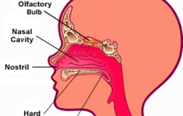
The human nasal cavity

The physical features in a person's face are the result of the way skin, fat and muscle tissue lie over this bony structure. An exception is the human nose. The external portion of the nose is mostly cartilage and connective tissue covered with skin. Hair and mucous line the nose and protect the interior nasal passageways from dust, debris and other foreign substances.

The external portion of the nose is a little deceptive. Two nostrils appear to lead almost straight up into the nasal passageway. In reality, the nasal cavity, which connects the nose to the throat, leads almost straight back. Its ceiling is approximately even with the top of the nose, just below the eyes. Its floor tends to be almost level with the alar cartilage, which forms the openings for the nostrils.

The nasal cavity isn't entirely smooth and straight. Its walls are made up of several grooves known as conchae. These grooves hold on to moisture when you exhale through your nose, which helps keep your nasal passages from drying out. Mucous membranes line all of these surfaces, providing lubrication and protection.

Diagram of the Human Nose

Anatomy and the Human Blockhead Video :