Structure, Function, and Clinical Significance
The human nose is far more than a simple airway for breathing. It is a complex, highly specialized organ responsible for respiration, olfaction (smell), immune defense, and speech resonance. Its intricate physiology allows the body to filter, warm, humidify, and analyze the air we breathe—while simultaneously protecting the lower respiratory tract from pathogens and irritants.
This article explores the anatomy and physiology of the human nose, how it functions in health, and why it is vital to overall respiratory and neurological well-being.
Overview: What Is the Human Nose?
Physiologically, the nose is the primary entrance to the respiratory system and the sensory organ for smell. It works continuously to:
-
Regulate airflow
-
Condition inhaled air
-
Detect odors
-
Provide immune defense
-
Assist speech resonance
The nose is divided into external and internal components, each contributing to its overall function.
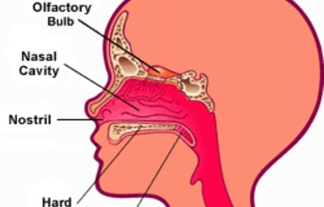
Anatomy of the Human Nose (Physiological Perspective)
1. External Nose
The external nose includes:
-
Nostrils (nares)
Physiological role:
-
Directs airflow
-
Provides structural support
-
Influences airflow resistance and turbulence
2. Nasal Cavity
The nasal cavity is divided into two chambers by the nasal septum and lined with specialized mucous membranes.
Key features:
-
Vestibule – initial air entry
-
Respiratory region – air conditioning
-
Olfactory region – smell detection
3. Nasal Turbinates (Conchae)
Three bony structures on each side:
Physiological functions:
-
Increase surface area
-
Create airflow turbulence
-
Enhance warming and humidification
-
Improve odor detection
Physiology of Nasal Breathing
1. Air Filtration
As air enters the nose:
-
Vibrissae (nose hairs) trap large particles
-
Mucus captures dust, bacteria, and allergens
-
Cilia move contaminants toward the throat for swallowing or expulsion
This process is part of the mucociliary clearance system, a key innate immune defense.
2. Air Warming and Humidification
The nasal lining is rich in blood vessels.
Physiological effects:
-
Warms cold air to near body temperature
-
Adds moisture to prevent airway dehydration
-
Protects lung tissue from irritation
Up to 90% of air conditioning occurs in the nose before air reaches the lungs.
3. Regulation of Airflow Resistance
The nose naturally regulates airflow through:
-
Nasal valve area
-
Turbinate swelling and decongestion cycles
This ensures optimal oxygen uptake and balanced airflow between nostrils.
Olfactory Physiology: How the Nose Enables Smell
Olfactory Epithelium
Located in the upper nasal cavity, it contains:
-
Olfactory receptor neurons
-
Supporting cells
-
Basal stem cells
Odor molecules dissolve in mucus and bind to receptors, triggering nerve impulses.
Signal Transmission
-
Odorant binds receptor
-
Electrical signal generated
-
Signal travels via olfactory nerve
-
Processed in olfactory bulb
-
Sent to brain regions for memory and emotion
This explains why smell is strongly linked to memory and emotion.

Immune and Protective Functions of the Nose
The nose is a frontline immune organ.
Defense Mechanisms Include:
-
Antimicrobial enzymes (lysozyme, defensins)
-
Immunoglobulin A (IgA)
-
Resident immune cells
-
Mucus barrier
These mechanisms help prevent infections such as:
Nasal Cycle: A Unique Physiological Phenomenon
The nasal cycle is an alternating congestion and decongestion of nostrils every 2–6 hours.
Purpose:
-
Allows tissue recovery
-
Maintains mucosal health
-
Optimizes air conditioning
This process is normal and usually unnoticed.
Role of the Nose in Speech and Resonance
The nose contributes to vocal quality by:
-
Acting as a resonating chamber
-
Modulating airflow during speech
-
Enabling nasal sounds (m, n, ng)
Blocked nasal airflow alters voice tone, resulting in hyponasal speech.
Clinical Relevance of Nasal Physiology
Disruption of nasal physiology can lead to:
-
Anosmia (loss of smell)
Understanding nasal physiology is critical for:
-
ENT diagnosis
-
Respiratory health
-
Surgical planning
-
Allergy management
The Nose and Whole-Body Health
Proper nasal function supports:
-
Lung efficiency
-
Brain oxygenation
-
Immune health
-
Sleep quality
-
Exercise performance
Mouth breathing bypasses these benefits and may negatively affect long-term health.
Conclusion
The physiology of the human nose reflects a remarkable balance of structure, function, and defense. Far beyond its role in breathing, the nose is a dynamic organ that conditions air, protects the body, enables smell, and contributes to communication and neurological processing.
Maintaining healthy nasal function is essential for optimal respiratory performance and overall well-being.