DIAGRAM OF THE HUMAN NOSE

UNDERSTANDING NASAL ANATOMY


 UNDERSTANDING NASAL ANATOMY

The Gateway to Respiratory and Olfactory Health

The human nose is more than a facial feature—it is a complex organ integral to respiration, smell, immunity, and speech. Its intricate anatomy allows the body to filter, warm, and humidify inhaled air while protecting the lungs from pathogens and irritants. Understanding nasal anatomy is key to appreciating how this remarkable organ supports overall health.

This comprehensive guide explores the structure of the nose, its functional regions, and clinical relevance.


External Nose: The Visible Gateway

The external nose forms the most recognizable part of the face and serves as the primary entrance for air.

Key Features:

  • Nasal bones: Form the upper bridge.

  • Cartilage: Shapes the tip and nostrils.

  • Nostrils (nares): Two openings allowing air entry.

Functionality:

  • Directs airflow efficiently into the nasal cavity.

  • Provides resistance to optimize breathing.

  • Supports the face structurally and aesthetically.


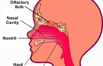
Internal Nasal Anatomy

The internal nose, or nasal cavity, extends from the nostrils to the nasopharynx and is divided by the nasal septum into two chambers.

1. Nasal Septum

  • Made of bone and cartilage.

  • Separates the two nostrils.

  • Provides structural stability and regulates airflow.

2. Nasal Cavity Lining

  • Covered with mucous membrane.

  • Houses cilia, which move mucus and trapped particles toward the throat.

  • Produces mucus to trap dust, bacteria, and allergens.


Nasal Conchae (Turbinates): The Air Conditioners

Three bony structures on each side of the nasal cavity:

  1. Inferior turbinate

  2. Middle turbinate

  3. Superior turbinate

Physiological roles:

  • Increase surface area for air contact.

  • Create turbulence to warm, humidify, and filter air.

  • Improve olfactory exposure for smell detection.


Olfactory Region: The Sense of Smell

Located in the upper nasal cavity, this region contains olfactory epithelium, consisting of:

Function:

  • Detects odor molecules dissolved in mucus.

  • Sends signals to the brain via the olfactory nerve.

  • Connects smell with memory and emotion, demonstrating the nose’s neurophysiological importance.


Nasal Sinuses: Lightweight and Resonant Structures

The paranasal sinuses—frontal, maxillary, ethmoid, and sphenoid—are air-filled cavities surrounding the nasal cavity.

Roles in nasal anatomy:

  • Reduce skull weight.

  • Produce mucus to trap pathogens.

  • Serve as resonating chambers for speech.


Blood Supply and Nervous Connections

  • Blood supply: Primarily from the sphenopalatine, facial, and ophthalmic arteries.

  • Innervation: Trigeminal nerve provides sensation; olfactory nerve transmits smell.

  • Rich vascularization contributes to air warming and plays a role in nosebleeds (epistaxis).


Nasal Physiology and Clinical Relevance

Understanding nasal anatomy explains common conditions:

  • Deviated septum: Alters airflow, may cause snoring or sleep apnea.

  • Nasal polyps: Obstruct airflow and sinus drainage.

  • Rhinitis and sinusitis: Result from inflammation of mucosa.

  • Anosmia (loss of smell): Often related to olfactory region damage or infection.

Proper nasal function ensures efficient breathing, optimal oxygen uptake, and immune defense.


The Nasal Cycle: An Anatomical Curiosity

The nasal cycle is a physiological alternation in congestion and decongestion between nostrils every few hours.
Purpose:

  • Allows mucosa recovery.

  • Maintains airway efficiency.

This subtle anatomical feature is normal and supports overall nasal health.


Conclusion

Understanding nasal anatomy is fundamental for appreciating how this small but complex organ impacts respiratory health, sensory perception, and overall well-being. From filtering and conditioning air to detecting odors and supporting speech, the nose is a marvel of anatomical design.

Maintaining nasal health through hygiene, proper hydration, and prompt treatment of infections helps preserve its critical physiological functions.

OTHER SOURCES


A lot happens under the surface of the nose. The bone and cartilage under the skin give the nose most of its size and shape. Other structures inside and behind the nose help you breathe. Learning the anatomy of the nose can help you better understand how the nose works.

Bone. 

This supports the bridge of the nose. The upper cartilage supports the side of the nose. The lower cartilage adds support, width, and height. It helps shape the nostrils and the tip of the nose.

Skin. 

This also helps shape the nose.

Nasal cavity. 

This is a hollow space behind the nose that air flows through.

Septum. 

This is a thin wall made of cartilage and bone. It divides the inside of the nose into 2 parts.

Mucous membrane. 

This is thin tissue that lines the nose, sinuses, and throat. It warms and moistens the air you breathe in. It also makes the sticky mucus that helps clean the air of dust and other small particles.

Turbinates. 

These are curved, bony ridges on each side of the nose. They are lined with mucous membrane. They warm and moisten the air you breathe in.

Sinuses. 

These are hollow, air-filled chambers in the bone around your nose. Mucus from the sinuses drains into the nasal cavity.

 UNDERSTANDING NASAL ANATOMY VIDEO :


Physiology of the Human Nose


Physiology of the Human Nose

Structure, Function, and Clinical Significance

The human nose is far more than a simple airway for breathing. It is a complex, highly specialized organ responsible for respiration, olfaction (smell), immune defense, and speech resonance. Its intricate physiology allows the body to filter, warm, humidify, and analyze the air we breathe—while simultaneously protecting the lower respiratory tract from pathogens and irritants.

This article explores the anatomy and physiology of the human nose, how it functions in health, and why it is vital to overall respiratory and neurological well-being.


Overview: What Is the Human Nose?

Physiologically, the nose is the primary entrance to the respiratory system and the sensory organ for smell. It works continuously to:

  • Regulate airflow

  • Condition inhaled air

  • Detect odors

  • Provide immune defense

  • Assist speech resonance

The nose is divided into external and internal components, each contributing to its overall function.


Anatomy of the Human Nose (Physiological Perspective)

1. External Nose

The external nose includes:

Physiological role:

  • Directs airflow

  • Provides structural support

  • Influences airflow resistance and turbulence


2. Nasal Cavity

The nasal cavity is divided into two chambers by the nasal septum and lined with specialized mucous membranes.

Key features:


3. Nasal Turbinates (Conchae)

Three bony structures on each side:

Physiological functions:

  • Increase surface area

  • Create airflow turbulence

  • Enhance warming and humidification

  • Improve odor detection


Physiology of Nasal Breathing

1. Air Filtration

As air enters the nose:

  • Vibrissae (nose hairs) trap large particles

  • Mucus captures dust, bacteria, and allergens

  • Cilia move contaminants toward the throat for swallowing or expulsion

This process is part of the mucociliary clearance system, a key innate immune defense.


2. Air Warming and Humidification

The nasal lining is rich in blood vessels.

Physiological effects:

  • Warms cold air to near body temperature

  • Adds moisture to prevent airway dehydration

  • Protects lung tissue from irritation

Up to 90% of air conditioning occurs in the nose before air reaches the lungs.


3. Regulation of Airflow Resistance

The nose naturally regulates airflow through:

  • Nasal valve area

  • Turbinate swelling and decongestion cycles

This ensures optimal oxygen uptake and balanced airflow between nostrils.


Olfactory Physiology: How the Nose Enables Smell

Olfactory Epithelium

Located in the upper nasal cavity, it contains:

  • Olfactory receptor neurons

  • Supporting cells

  • Basal stem cells

Odor molecules dissolve in mucus and bind to receptors, triggering nerve impulses.


Signal Transmission

  1. Odorant binds receptor

  2. Electrical signal generated

  3. Signal travels via olfactory nerve

  4. Processed in olfactory bulb

  5. Sent to brain regions for memory and emotion

This explains why smell is strongly linked to memory and emotion.


Immune and Protective Functions of the Nose

The nose is a frontline immune organ.

Defense Mechanisms Include:

  • Antimicrobial enzymes (lysozyme, defensins)

  • Immunoglobulin A (IgA)

  • Resident immune cells

  • Mucus barrier

These mechanisms help prevent infections such as:


Nasal Cycle: A Unique Physiological Phenomenon

The nasal cycle is an alternating congestion and decongestion of nostrils every 2–6 hours.

Purpose:

  • Allows tissue recovery

  • Maintains mucosal health

  • Optimizes air conditioning

This process is normal and usually unnoticed.


Role of the Nose in Speech and Resonance

The nose contributes to vocal quality by:

  • Acting as a resonating chamber

  • Modulating airflow during speech

  • Enabling nasal sounds (m, n, ng)

Blocked nasal airflow alters voice tone, resulting in hyponasal speech.


Clinical Relevance of Nasal Physiology

Disruption of nasal physiology can lead to:

Understanding nasal physiology is critical for:

  • ENT diagnosis

  • Respiratory health

  • Surgical planning

  • Allergy management


The Nose and Whole-Body Health

Proper nasal function supports:

  • Lung efficiency

  • Brain oxygenation

  • Immune health

  • Sleep quality

  • Exercise performance

Mouth breathing bypasses these benefits and may negatively affect long-term health.


Conclusion

The physiology of the human nose reflects a remarkable balance of structure, function, and defense. Far beyond its role in breathing, the nose is a dynamic organ that conditions air, protects the body, enables smell, and contributes to communication and neurological processing.

Maintaining healthy nasal function is essential for optimal respiratory performance and overall well-being.


OTHER SOURCES

THE HUMAN NOSE

The human nose has several physiologic functions. As the air is inspired through the nose it is humidified and warmed by passing over the moist and warm nasal mucosa. The nose is an energy-conscious organ in that expired air is cooled and some of the moisture is recaptured. The functions of warming and humidification require a tremendous blood flow to the nasal mucosa and also place substantial stress on the nasal mucosa. The nose has what is known as a nasal cycle. 

To allow the mucosa time to rest from these functions, the nose has periods in which one side of the nose becomes swollen, which we call congestion. During the same period the other side of the nose remains patent or decongested. The normal nasal cycle lasts from three to six hours, and during this time first one side of the human nose will be congested and the other side will be patent; then the second side becomes congested as the first side becomes patent. It is also normal for the human nose to be congested when an individual lies down.

Particularly if you lie on your side you may notice that the downside of your nose becomes congested. Olfaction is the medical word for smell. Smell is one of the six human senses and for some individuals is a very important sense; for others it seems to be less important. 

The olfactory centers, as described under the anatomy section, reside very high along the roof of the nose. In order for these centers to be stimulated, the odors have to be inspired into the human nose and carried up to the roof of the human nose. If the odor does not reach the roof of the nose due to a variety of conditions, the odor will not be perceived. The nose is capable of distinguishing a large number of different odors. Our understanding of precisely how this occurs is limited, but increasing.

Taste is a sense very different from olfaction. It is another of the human senses and is perceived primarily on the tongue. There are four recognized tastes and these are sweet, salty, sour, and bitter. They occupy geographically separate areas on the tongue and are perceived in cells clustered together in taste buds. The sense of smell plays a major role in the flavor of foods and it is common for individuals who lose their sense of smell to report that food loses its taste. This is of course incorrect; the food has only lost its aroma, and taste (sweet, salty, sour, bitter) remains intact.

Physiology of the Human Nose Video :\


The Anatomy of the Nose


The Anatomy of the Nose

The nose is the part of the respiratory tract that sits front and center on your face. You use it to breathe air in and to stop and smell the roses. The nose’s exterior anatomy includes the nasal cavity, paranasal sinuses, nerves, blood supply, and lymphatics.

The external part of the nose includes the root (between the eyes), the dorsum that runs down the middle, and the apex at the tip of the nose. Two openings called nostrils (nares) allow air in. They’re divided by the nasal septum (dividing wall of cartilage and bone), and the parts that surround the nostrils are called the alae (ala singular).

The nose has a bony part that’s formed by the bony nasal septum, the nasal bones, and parts of the maxillae, palatine, and frontal bones. The cartilaginous part of the nose is formed by two lateral cartilages, two alar cartilages, and a septal cartilage.

The nasal cavity
The nares serve as the entryway to the nasal cavities, which open posteriorly into the nasopharynx via the choanae. The walls of the nasal cavity include the following features:

Roof: The roof is divided into three parts: frontonasal, ethmoidal, and sphenoidal. Each part corresponds to the underlying bone of the same name.

Floor: The floor consists of the palatine process of the maxilla and the horizontal plate of the palatine bone.

Medial wall: This wall is the nasal septum, which is formed by the perpendicular plate of the ethmoid bone, the vomer, cartilage, and the nasal crests of the maxillary and palatine bones.

Lateral wall: This wall is hallmarked by three nasal conchae (superior, middle, and inferior) that project inferiorly from the wall. They divide the nasal cavity into four passages that have openings to the paranasal sinuses:

The sphenoethmoid recess lies posterior to the superior concha and has the opening for the sphenoidal sinus.

The superior nasal meatus lies between the superior and middle conchae and has openings to the posterior ethmoidal sinuses.

The middle nasal meatus is longer and deeper than the superior nasal meatus. The frontal sinus communicates with the middle nasal meatus via the infundibulum, a passageway that opens into the semilunar hiatus (groove in the ethmoid bone). The maxillary sinus opens into the semilunar hiatus. An ethmoidal bulla (a round swelling formed by the middle ethmoidal cells, or air-filled cavities) is formed just above the semilunar hiatus. The middle and anterior ethmoidal sinuses drain into the middle nasal meatus.

The inferior nasal meatus is found below the inferior nasal concha. The nasolacrimal duct opens into this meatus.

The nasal cavity is lined with nasal mucosa, except for the nasal vestibule, which is lined with skin. The mucosa over the superior one-third of the nasal cavity is the olfactory area. Air is drawn past the specialized mucosal cells called the olfactory epithelium as air is sniffed though the nose. 

The olfactory epithelium contains receptors of olfactory neurons that detect smells. Olfactory neurons (from CN I) join together to form nerve bundles that run up through the cribiform plate of the ethmoid bone to the olfactory bulb. The olfactory tract transmits the sensory information about smell from

The paranasal sinuses
The paranasal sinuses are air-filled cavities in the frontal, ethmoid, maxilla, and sphenoid bones. They’re lined with a mucosal membrane and have small openings into the nasal cavity:

Maxillary sinus: This sinus is located in the body of the maxilla behind the cheek just above the roots of the premolar and molar teeth. It’s shaped like a pyramid. It opens into the nasal cavity via the semilunar hiatus.

Frontal sinuses: Found within the frontal bone, each of these sinuses is triangular in shape and runs above the medial end of the eyebrow and backward to the orbit. They open into the nasal cavity via the semilunar hiatus.

Sphenoid sinuses: These sinuses are found in the sphenoid bone. Each opens into the sphenoethmoid recess.

Ethmoid sinuses: The anterior, middle, and posterior ethmoid sinuses are located in the ethmoid bone between the nose and the eye. The anterior sinus opens into the nasal cavity by the infundibulum, the middle sinus opens into the ethmoidal bulla, and the posterior sinus opens into the superior meatus.

Nerves, blood vessels, and lymphatics of the nose

Nerve supply to the external nose is provided by the infratrochlear and external nasal branches of the ophthalmic nerve and the infraorbital branch of the maxillary nerve, both of which are part of the trigeminal nerve (CN V). 
The olfactory nerves (CN I) pass through the cribiform plate of the ethmoid bone. General sensory innervation of the nasal cavity and the paranasal sinuses is from the ophthalmic nerve (CN V1) and maxillary nerve (CN V2).

Blood is supplied to the external part of the nose by branches of the ophthalmic and maxillary arteries. The skin of the ala and septum are supplied by the facial artery. 

Blood is brought to the walls of the nasal cavity and sinuses by branches of the maxillary artery. The most important is the sphenopalatine artery, which anastomoses with a branch of the superior labial artery. Venous blood is returned from the nasal cavity by veins that accompany the arteries.

Lymph from the nasal cavity drains into the submandibular lymph nodes and vessels that drain into the upper deep cervical lymph nodes.
The Anatomy of the Nose Video :


Anatomy and the Human Blockhead


Anatomy and the Human Blockhead

🧠 The Anatomy Behind the Illusion

1. The Nasal Cavity Is Deeper Than It Looks

Inside the nose is a long, air-filled chamber that extends straight back toward the throat. When you look at someone’s nostrils from outside, it seems like the space ends quickly—but it actually continues much further back in a straight line.

  • The average adult nasal cavity is 7–8 cm (about 3 inches) deep.

  • It’s shaped like a tunnel, not an upward turn the way people often imagine.

This is the space a performer uses.


2. What the Performer Is Not Doing

  • They are NOT piercing bone.

  • They are NOT entering the brain (the “brain through the nose” myth).

  • They are NOT going anywhere near the skull plate between the nasal cavity and the brain.

The nail simply follows the natural path of the nasal passage.


3. Why It Looks So Dramatic

  • Most people assume the nose turns upward right away, so the straight-back direction feels wrong.

  • The object is long, rigid, and looks like it must be going into solid bone, but it isn’t.

  • The performer’s facial expression (remaining calm) enhances the illusion that something impossible is happening.


4. Why Performers Can Do It Safely (After Training)

Professional performers:

  • Understand nasal anatomy precisely.

  • Know how to avoid delicate structures like blood vessels and mucous membranes.

  • Build tolerance over time.

  • Use sterile objects and proper hygiene.

  • Know how to stop if they hit resistance or irritation.


⚠️ Important Safety Note

Although the stunt is based on real anatomy, it is not safe for untrained individuals. Attempting it can cause:

Professional sideshow performers train for months or years under experienced supervision.


OTHER SOURCES

THE HUMAN NOSE

The human blockhead act is a trick that relies on the structure of the human skull

The skull is a collection of 22 bones, most of which are fused together at joints called sutures

These fused bones form a covering that protects your brain. Although much of the skull looks like one solid piece, it's also full of holes and spaces. 

These include the sinuses, the eye sockets and the foramen magnum, where the brain stem exits the skull.

The human nasal cavity

The physical features in a person's face are the result of the way skin, fat and muscle tissue lie over this bony structure. An exception is the human nose. The external portion of the nose is mostly cartilage and connective tissue covered with skin. Hair and mucous line the nose and protect the interior nasal passageways from dust, debris and other foreign substances.

The external portion of the nose is a little deceptive. Two nostrils appear to lead almost straight up into the nasal passageway. In reality, the nasal cavity, which connects the nose to the throat, leads almost straight back. Its ceiling is approximately even with the top of the nose, just below the eyes. Its floor tends to be almost level with the alar cartilage, which forms the openings for the nostrils.

The nasal cavity isn't entirely smooth and straight. Its walls are made up of several grooves known as conchae. These grooves hold on to moisture when you exhale through your nose, which helps keep your nasal passages from drying out. Mucous membranes line all of these surfaces, providing lubrication and protection.

Diagram of the Human Nose

Anatomy and the Human Blockhead Video :


Anatomy and Physiology of the Nose and Throat


Anatomy and Physiology of the Nose and Throat

What is the human nose?

The human nose is the organ of smell located in the middle of the face. The internal part of the nose lies above the roof of the mouth. The human nose consists of:

  • external meatus - triangular-shaped projection in the center of the face.
  • external nostrils - two chambers divided by the septum.
  • septum - made up primarily of cartilage and bone and covered by mucous membranes. The cartilage also gives shape and support to the outer part of the nose.
  • nasal passages - passages that are lined with mucous membranes and tiny hairs (cilia) that help to filter the air.
  • sinuses - four pairs of air-filled cavities, also lined with mucous membranes.

What are sinuses?

The sinuses are cavities, or air-filled pockets, near the nasal passage. As in the nasal passage, the sinuses are lined with mucous membranes. There are four different types of sinuses:

  • ethmoid sinus - located inside the face, around the area of the bridge of the nose. This sinus is present at birth, and continues to grow.
  • maxillary sinus - located inside the face, around the area of the cheeks. This sinus is also present at birth, and continues to grow.
  • frontal sinus - located inside the face, in the area of the forehead. This sinus does not develop until around 7 years of age.
  • sphenoid sinus - located deep in the face, behind the nose. This sinus does not develop until adolescence.
 What is the throat?

The throat is a ring-like muscular tube that acts as the passageway for air, food, and liquid. The throat also helps in forming speech. The throat consists of:

  • larynx - also known as the voice box, the larynx is a cylindrical grouping of cartilage, muscles, and soft tissue which contains the vocal cords. The vocal cords are the upper opening into the windpipe (trachea), the passageway to the lungs.
  • epiglottis - a flap of soft tissue located just above the vocal cords. The epiglottis folds down over the vocal cords to prevent food and irritants from entering the lungs.
  • tonsils and adenoids - made up of lymph tissue and are located at the back and the sides of the mouth. They protect against infection, but generally have little purpose beyond childhood.
Diagram of the Human Nose , Anatomy and Physiology of the Nose and Throat : 


The Nose


The Nose

1. Overview

The nose is the primary organ for breathing and smelling. It also filters, warms, and humidifies the air before it reaches the lungs. It is divided into:

  • External nose – the visible part on the face.

  • Internal nose (nasal cavity) – the hollow space inside.


2. External Nose

  • Nasal bridge: The bony upper part of the nose.

  • Nostrils (nares): Openings for air entry.

  • Nasal tip: The protruding end of the nose.

  • Ala: The rounded flared sides of the nostrils.

  • Columella: The tissue separating the two nostrils.


3. Internal Nose (Nasal Cavity)

  • Nasal septum: Divides the nasal cavity into right and left sides; made of bone and cartilage.

  • Nasal conchae (turbinates): Superior, middle, inferior – bony curves that increase surface area for air filtration, warming, and humidification.

  • Meatuses: Air passages beneath each concha.

  • Olfactory epithelium: Located at the top; contains smell receptors.

  • Respiratory epithelium: Lining most of the cavity; secretes mucus to trap particles.


4. Sinuses (Paranasal Sinuses)

Hollow spaces connected to the nasal cavity:

  • Frontal sinus – above the eyes.

  • Maxillary sinus – in the cheeks.

  • Ethmoid sinus – between the eyes.

  • Sphenoid sinus – deep behind the ethmoid sinuses.


5. Functions

  1. Breathing: Passageway for air to reach lungs.

  2. Smelling: Houses olfactory receptors.

  3. Air conditioning: Warms, humidifies, and filters inhaled air.

  4. Resonance: Contributes to voice sound.

  5. Protection: Traps dust, microbes, and other particles with mucus and cilia.


The nose is the body's primary organ of smell and also functions as part of the body's respiratory system.

Air comes into the body through the nose. As it passes over the specialized cells of the olfactory system, the brain recognizes and identifies smells. Hairs in the nose clean the air of foreign particles. As air moves through the nasal passages, it is warmed and humidified before it goes into the lungs.

The most common medical condition related to the nose is nasal congestion. This can be caused by colds or flu, allergies, or environmental factors, resulting in inflammation of the nasal passages. The body's response to congestion is to convulsively expel air through the nose by a sneeze.

Nosebleeds, known medically as epistaxis, are a second common medical issue of the nose. As many as 60 percent of people report nosebleed experiences, with the highest rates found in children under 10 and adults over 50.


diagram-of-human-nose.

DIAGRAM OF THE HUMAN NOSE-3


DIAGRAM OF THE HUMAN NOSE-3

Here’s a clear textual breakdown of a diagram of the human nose (I can also generate an actual labeled image if you want):

1. External Nose (visible part):

  • Nasal bridge: The upper bony part of the nose.

  • Nostrils (nares): Openings for air entry.

  • Nasal tip: The pointed end of the nose.

  • Ala (plural: alae): The flared sides around the nostrils.

  • Columella: Tissue that separates the two nostrils.

2. Nasal Cavity (internal):

  • Nasal septum: Divides the nasal cavity into right and left sides; made of cartilage and bone.

  • Nasal conchae (turbinates): Three curved bony structures (superior, middle, inferior) that increase surface area for warming and humidifying air.

  • Nasal meatuses: Passages beneath each concha for air flow.

  • Olfactory epithelium: Located at the top of the nasal cavity; responsible for smell.

  • Respiratory epithelium: Lines most of the nasal cavity; contains mucus-secreting cells.

3. Sinuses (connected to nasal cavity):

4. Nasopharynx (back of nasal cavity):

VIDEO: